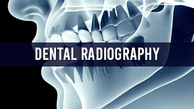
Dental radiographs are generally called X-rays. Radiographs are used for many reasons: bone loss, to find hidden dental structures, cavities, malignant or/and benign masses. By a controlled burst of X-ray radiation a radiographic image is formed. This ray inserts oral structures at various levels, depending on different anatomical densities, before reaching the film or sensor. Changes in the bone density, dental caries, dental infections and the periodontal ligament, appear darker because X-rays readily penetrate these less dense structures and teeth appear lighter because less radiation penetrates them to reach the film. Dental restorations (fillings, crowns) may appear lighter or darker, depending on the density level of the material.
Sub-tracks:
• Ionizing radiation
• Gamma rays
• Radiographic film
Share this page on your timeline
ALSO READ Orthodontics Prosthodontics Endodontics Oral Microbiology Dental Anesthesiology 3d Imaging and Digital Dentistry Dental Sleep Medicine Oral Cancer Holistic Dentistry Pediatric Dentistry Pregnancy and Oral Health Dental Surgery Dental Materials Dental Nursing General Dental Council Dental Robotics Dentures Cosmetic Dentistry Dental Traumatology Dental Radiography Laser Dentistry Nano Dentistry Restorative Dentistry Forensic Odontology Current Concept on Oral Health Dental Implant Dental Anatomy DENTAL CARIES Dental Education Geriatric Dentistry Veterinary Dentistry Dentists Dental Public Health Dental Students
Tags
Oral Biology Conferences
Dental Conferences 2021 USA
Dental Conferences 2021 Europe
Cosmetic Dentistry Conferences
Dental Conferences 2021 Asia
Dental Restoration Conferences
Dental Conferences
Dental Medicine Conferences
Endodontics Conferences
Dental surgery Conferences
Holistic Dentistry Conferences
Dental Conferences 2021 Canada
Restorative Dentistry Conferences
Dentist Conferences
Restorative Dentistry Conferences